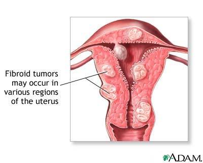
Uterine fibroids are BENIGN i.e.
non-cancerous), usually clusters of TUMORS in, on or
within the uterine walls
Originate from and
are composed of SMOOTH MUSCLE cells(myocytes)
of the
uterine wall's muscle layer (called the myometrium, the middle layer of the uterine wall used for
contracting the uterus) and
its accompanying connective tissue;
It is RARE for benign uterine
leiomyomas to progress to cancerous leiomyosarcomas (1.7 women per 100,000 women are diagnosed annually with
uterine sarcoma, which includes leiomyosarcoma)
(National Cancer Institute)
Vary in size - from microscopic to very large (can weigh several pounds).
Fibroids are often described by their location in
the uterus:
Myometrial - in
the muscle wall of the uterus
Submucosal -
just under the surface of the uterine lining
Subserosal
- just under the outside covering of the uterus
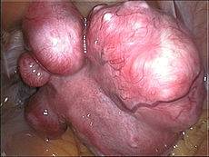
Pendunculated -
occurring on a long stalk on the outside of the uterus or inside the cavity of
the uterus
Called DIFFUSE uterine leiomyomatosis - when there are too
many fibroids to count.
Diagnosis of uterine fibroids
Diagnosis can be wrong - pelvic
examination may show an irregularly shaped, lumpy, or enlarged uterus, but in
obese women it is difficult to diagnose fibroids, which may be mistaken for:
Pregnancy
Ovarian tumors
Inflammation of the fallopian tubes
Uterine adenomyosis (a
condition in which the uterine lining grows into the muscle wall of the uterus)
Fibroids can be confirmed - by a transvaginal ultrasound, a pelvic
ultrasound or a pelvic MRI.
Who gets uterine fibroids?
UFs are the most common pelvic tumor in females,
typically found during the mid- to late-reproductive years
Uterine fibroid incidence
rate is 70% by age 50 in U.S. white women and 80% in African-American women -
with a staggering half of reproductive age U.S. women having fibroids. Typically affects women over 30, but not under 20;
(Baird et al, 2003)
More common in
African-American women than Caucasian
women - ~25% of white women and 50% of black women have
symptomatic uterine fibroids. (Wise,
2005)
More common in overweight
women -perhaps
because of increased
estrogenfrom adipose aromatase
enzyme
activity that converts the androgens
ANDROSTENEDIONE and
Testosterone to
estrogens
UFs often have a
growth spurt before menopause -and then become
quiescent
Higher UF incidence in women who have not given
birth or had early menarche or have a UF history in first degree relatives
Smokers have LESS
risk of UFs
Physical activity
seems to protect against having UFs (Baird
et al, 2007) (Flake et al, 2003)
What are the symptoms of
uterine fibroids?
Not all UFs are symptomatic, but those that are can grow,
causing:
Heavy and painful menstruation; bleeding
between periods; longer-lasing periods
Painful sexual intercourse
Urinary frequency and urgency
Pelvic pain / pressure;
Abdominal fullness, gas, constipation
Pregnancy complications (rare) -
increased blood flow and estrogen levels during pregnancy may cause UFs to grow,
but return to normal size after delivery. Insufficient room in uterus may
require early delivery; C-section may be needed if UFs block birth canal or
cause wrong positioning of baby; may cause heavy bleeding immediately after
giving birth.
Other complications of fibroids include:
A pedunculated fibroid can become twisted and
cause a kink in the blood vessels feeding the tumor - may need surgery;
Anemia - may be
severe with heavy bleeding
Urinary tract infections -
pressure from the fibroid can prevent bladder emptying fully;
Malignant change
(extremely rare) - called a leiomyosarcoma
Infertility
(rare)
What causes uterine fibroids?
Hormonal influences and growth factors are involved in
UF growth and development:
During a woman's menstruating years, UFs
typically continue to grow slowly
Large fibroids may outgrow their blood supply and
degenerate - described as hyaline, myxomatous, calcific, cystic, fatty, necrotic or red
(usually only during pregnancy).
Fibroid growth seems to depend on both
estrogen and Progesteronehormones
UFs have excessive production of a disorganized but stable extracellular matrix
(ECM) and altered collagen fibrils
in the ECM - fibroid collagen fibers (bunches of fibrils) are short,
widely dispersed and lying non-parallel, compared to well-packed and
lying parallel in the myometrium (smooth muscle tissue of the uterus).
It is the abnormal and overproduced ECM that causes UF expansion ,
and not the slowly proliferating fibroid cells - UF tumors contain decreased/disrupted matrix metalloproteinases (MMPs)
and more proteins in their ECM, such as collagen subtypes, proteoglycans,
fibronectin, matrix glycoproteins and matricellular proteins (in particular
thrombospondin-1 (TSP-1), which activates
TGF- β and has a role
in angiogenesis). The ECM binds cytokines and growth factors ready for action in
the vicinity of the UF. Integrins are changed in UFs. The stability of this allbeit disorganized ECM
requires therapeutic interventions that address ECM dissolution in addition to
inhibiting cell proliferation
UFs involve growth factors:
Transforming Growth
Factors-β1 and β3 (TGF- β1,TGF- β3) - have a central role in UF enlargement, in that they stimulate
production/deposition of ECM and are acknowledged as important growth factors in
fibrotic disease. E.g. Fibroids have more concentrated TGF-β receptors.
Conversely, reduced TGF-β expression
yields reduced ECM production and fibroid shrinkage
Increased profibrotic cytokines
(E.g. IL-1, IL-6, interferon, TNF- α)
in UFs
-
involved with inflammatory response, cytokines are produced when growth
factors act on target tissue.
UFs grow at different rates (even in the same
woman) - and with different growth-rate patterns in white and African-American
women
>50% of UFs are asymptomatic(i.e. have no symptoms) -
~70% of women by age 45 will be diagnosed
with UFs, but only a fraction of those
will cause problems or require treatment. (Merck Manual)
Organochlorine pesticides stimulate
leiomyomata cell proliferation in animals - organochlorines are
xenoestrogens (i.e. mimic
estrogen
in the body). (Hodges et al, 2000)
Hodges LC, Bergerson JS, Hunter DS, Walker CL. (2000 Apr) Estrogenic effects of
organochlorine pesticides on uterine leiomyoma cells in vitro. Toxicol Sci. ;54(2):355-364.
PubMed
Walker CL, Stewart EA (June 10, 2005) Uterine fibroids: the elephant in the room. Science. ; 308.
Estrogen
and
Progesterone generally promote uterine
fibroid growth
It's the "free" hormone levels that count. Some researchers
maintain that serum
estrogen
and
Progesterone levels are unchanged by
UFs - however, these serum levels are only
meaningful if their free levels
have been measured, which is not usually the case.
Overall effects of
Estrogenand
Progesterone:
Mitogenic effect on
leiomyoma cells. Encourages
cell division/mitosis;
Act by influencing (directly and indirectly)
a large
number of growth factors. Usually a protein or steroid hormone capable of stimulating
cellular growth, proliferation and cellular differentiation (less specialized
cell becomes a more specialized cell type).
EstrogenDominance.
A dominance of
estrogenover other hormones
is a recognized problem of today, due to dietary and environmental changes.
Fibroid cells can
make their ownESTRADIOLand the conversion enzymes to make it
are over-expressed in fibroids - Fibroids express
higher levels ofaromatase
and
can convert circulating
androstenedioneintoESTRADIOLvia the enzymesAromatase and
17ß-hydroxysteroid dehydrogenase (Walker
& Stewart, 2005;
Shozu, 2004)
Aromatase over-expression in uterine leiomyoma
tissue is particularly pronounced in African-American women
(Ishikawa et al, 2009)
LEPTIN (the "appetite suppressor" hormone) has also been shown to
increase
aromatase expression
Fibroids have a lot of
Estrogen receptors
Fibroid cells have moreestrogenreceptors (to
respond to estrogen)than normal uterine muscle cells
Having estrogen receptors, fibroids tend to enlarge during the
reproductive years and shrink after menopause -
In PREmenopausal fibroids the ER-ß,
ER-α (and
Progesterone) receptors are found
over-expressed -
compared to only
ER-ß in POSTmenopausal fibroids (which are rare)
(Strissel et al, 2007)
A special ER-αgenotype was found correlated with incidence and
size of fibroids -
Higher prevalence of this genotype in black women may also explain higher
incidence of fibroids in Afro-American women. Most studies found that other
different phenotypes in
ER and
Pr gene encodings are not correlated with
incidence of fibroids in Caucasian populations (Alhendy, 2006)
It is proposed that
Estrogen is
growth-promoting by up-regulating:
IGF-1,
EGFR,
TGF-β1 - Expression of transforming growth
interacting factor (TGIF) is increased in
leiomyoma compared with myometrium (In myometrial cells,TGIF is a potential
repressor of anti-proliferative TGF-β pathways).
Cytokines - signaling molecules secreted by nervous system glial cells and
many immune system cells for intercellular communication.
Apoptotic factors -
TGF-ß3 and
PDGF, promotes aberrant survival of leiomyoma cells by down-regulating the
tumor-suppressor protein p53.
Other hormones
Effects of Progesterone
on uterine fibroid growth
Uterine fibroids have more Progesterone receptors (to respond to
Progesterone) than normal uterine muscle cells.
Progesterone is thought to
promote the growth
of leiomyoma via up-regulation of
EGF,
TGF-β1 and TGF-β3
Progesterone is
thought to promote the survival
of leiomyoma via up-regulation of
Bcl-2
expression and down-regulating
TNF-α.
Progesterone is thought to counteract growth
of leiomyoma by downregulating IGF-1.
A recent study emphasized the anomaly whereby
>72% of women who were pregnant (or recently postpartum) have > 50% regression
of pre-existing fibroids - One explanation points to the postpartum fall
of Progesterone.
Progesterone
seems to have a dominant role by
INCREASING mitotic rates in fibroids in the
luteal phase of the menstrual cycyle (2nd
half of cycle when corpus luteum secretes a lot of Progesterone)-
the drug mifepristone, a
Progesterone antagonist,
INHIBITS fibroid growth lending support to Progesterone's dominant role. One theory is
that Progesterone upregulates EGF and TGF-β expression. However,
Progesterone also REDUCES the growth
factor IGF-1 in vitro and INHIBITS MMPs, which activate growth factors and
degrade extra cellular matrix (ECM), affecting ECM assembly and deposition, and
so counters UF enlargement.
Other notes
Actions ofestrogen
and Progesterone are
modulated by the "cross-talk" between themselves and
PROLACTIN - which controls the expression of their
respective nuclear receptors.
Rarely, leiomyomas progress to leiomyosarcomas
and evolve to a hormone-non-responsive state - since
many sarcomas have markedly reduced or
no steroid hormone receptors
Alhendy, A.; Salama, S.
(2006). "Ethnic distribution of ESTROGEN receptor-αpolymorphism is associated
with a higher prevalence of uterine leiomyomas in black Americans". Fertility
and Sterility 86 (3): 686
PubMed
Shozu,
M.; Murakami, K.; Inoue, M. (2004). "Aromatase and Leiomyoma of the Uterus".
Seminars in Reproductive Medicine 22 (1): 51.
PubMed
Strissel, P.; Swiatek, J.; Oppelt, P.; Renner, S.; Beckmann, M.; Strick, R.
(2007). "Transcriptional analysis of steroid hormone receptors in smooth muscle
uterine leiomyoma tumors of postmenopausal patients". The Journal of Steroid
Biochemistry and Molecular Biology 107(1-2): 42-47. .
PubMed
Mainstream treatments for symptomatic uterine fibroids
Oral contraceptives - to help control heavy periods
Intrauterine devices (IUDs)
that release the
synthetic hormone progestin - to help reduce heavy bleeding and pain
Iron supplements - to prevent or treat anemia due to heavy periods
Nonsteroidal anti-inflammatory drugs (NSAIDs) - E.g.ibuprofen for cramps or pain
Surgical Treatments
A
hysterectomy is frequently advised in the U.S. - especially
if a woman does not intend to have children. In fact, leiomyoma are the
predominant reason for a hysterectomy in premenopausal women
(MerckManual)
Myomectomy - This surgery removes the fibroids. It is often the chosen
treatment for women who want to have children, because it usually can preserve
fertility. More fibroids can develop after a myomectomy.
Magnetic Resonance-Guided Focused Ultrasound -Magnetic Resonance
guided Focused Ultrasound (MRgFUS), is a
non-invasive intervention (requiring no incision) that uses high intensity
focused ultrasound (HIFU) waves to ablate (destroy) tissue in combination with
Magnetic Resonance Imaging (MRI), which guides and monitors the treatment.
Hysteroscopic resection
of fibroids(as outpatient) - when UFs are growing inside the uterus. A small
camera/instruments are inserted through the cervix into the uterus to remove the
UFs.
Uterine artery embolization
- procedure cuts off blood supply to the UF, causing it to die and
shrink.
Anti-fibrotic therapies inhibit and reverse the fibrotic
process
Affect a change in abnormal ECM by leiomyoma cells
Aromatase inhibitors used to
reduce fibroids (Malartic, 2008)
- The effect is believed to be partially due to
(i) Lowering ovarian production and systemic
estrogen levels
and (ii) Inhibiting locally
overexpressed aromatase in fibroids.
Aromatase inhibitors have also been used
experimentally in treatment of
endometriosis - which indicated that aromatase inhibitors
might be particularly useful in combination with a progestogenic ovulation
inhibitor.
Phytoestrogens - compete for receptors with endogenous estrogens; isoflavones
daidzein and genistein are found in soy, but have been found to worsen fibroids
when consumed in too high amounts. Lignans found in flaxseed.
I3C in cruciferous vegetables - promotes formation of less potent estrogen
metabolites (Minich & Bland, 2007)
Reduce caffeine intake - to <500 mg / day
(e.g. 2 cups coffee / day)
Reduce alcohol consumption to 1 drink / day
Increase fiber - helps
remove excess estrogen from GI tract aiding
excretion; reduces enterohepatic estrogen recirculation and/or shields estrogen
absorption;
Consume anti-inflammatoryomega-3 fats and reduce inflammatory
omega-6 fats to reduceestrogen
production
Iodine
has a critical role in
maintaining the body's estrogenbalance and
can reduce uterine fibroids. Based on a controlled
clinical trial with 1,365 women, 4mg daily of molecular iodine quickly
resolves fibrocystic breast disease(FBD) -
Iodine makes breast lumps and cysts disappear usually within only two months
for most women.
Iodine
can similarly reduce uterine fibroids - one of the first conventional medical
treatments for severe fibroids was to "paint"the uterus with iodine.
The primary aromatasepromoter in leiomyomata tissues in non-Asian U.S. women is the inflammatory
prostaglandin PGE2 (Imir et al, 2007)
Omega-3fat
reduces release of growth hormone - which promotes formation /
growth of fibroids
Myoma is associated with beef and ham consumption,
whereas high intake of green vegetables seems to have a protective effect
(Chiaffarino et al, 1999)
Vitamin D3 decreases fibroid
cell size and disrupts the formation of fibroid muscle cells
Vitamin D3is typically deficient in many populations today - E.g.
Elderly, office workers, African
Americans
D3 treatment
has been shown to inhibit leiomyocyte proliferation at physiological doses -
Leiomyomas widely express the
vitamin D receptor.
Vitamin D
decreases mitogenic activity of INSULIN and
IGF-1
Active metabolites of CALCITRIOL (Active form of Vitamin D) down-regulate epidermal growth
factor receptors (EGFRs) known to be active in mitogenic pathways in uterine
leiomyomas. Down-regulation of these receptors shown to decrease growth /
differentiation of tumor cells
Risk of
developing uterine fibroids in American black women REDUCED with just increased daily servings of "vitamin
D-added" milk.
Research shows that physiological doses of
vitamin D have significant growth-inhibiting
effect on leiomyomata cells (Blauer et al, 2009)
There is an inverse association between bioflavonoid intake and
risk of malignant tumors -
reported biological
activities include:
Induce apoptosis
Cell cycle arrest
Antiproliferative
Anti-inflammatory
Antioxidant protection against
oxidative stress
Anti-estrogenic
Asian women consume a lot of bioflavonoids and
have lower incidence of hormonally dependent solid tumors - E.g. breast cancer in Asian women is 4-6
times lower than in American women, and several generations after migration to
America they line up with the American statistics, suggesting an environmental
rather than a genetic influence. Asian women consume a lot of soy-based foods,
containing bioflavonoids that show up in blood and urine samples at
significantly elevated levels.
"Bioflavonoids are . . . found
in legumes, nuts, onions, apple, broccoli, red wine, grreen tea, cocoa powder,
and dark chocolate. The best known anti-tumor flavonoids are epigallocatechin
gallate (EGCG)
from green tea, genistein (from soy and red clover),
curcumin (from
turmeric), silibin (from milk
thistle), quercetin (from many
yellow vegetables such as onions), and resveratrol (from grapes and red wine)."
Quercetin, EGCG, Curcumin, Silibrinin - (In berries, tea,
grapes, olive oil, dark chocolate, walnuts, citrus):
Inhibits IGF-1 signaling
Anti-estrogenic
-
Estrogen receptor antagonist
Alters cell cycle
Resveratrol (In red wine, grape, berries, dark
chocolate, also
peanuts (not recommended because of common fungal content); produced in plants in response to injury or fungal/
bacterial presence)
Curcumin
(spice) -
decreases growth and increases death of fibroid cells in vitro.
Curcumin inhibited uterine leiomyoma cell proliferation by inducing
apoptosis, and inhibited production of the ECM component fibronectin.
(Malik et al, 2009; Kenji et al, 2011; )
Licorice (contains flavonoid isoliquiritigenin) - decreases growth and increases apoptosis of fibroid cells
in vitro.
Green Tea (epigallocatechin gallate) - decreases growth of fibroid cells
in vitro.
Retinoic acid
GI tract health
strongly linked to uterine fibroid growth
Gastrointestinal problems (e.g. leaky gut syndrome, candida
(yeast), intestinal bacterial overgrowth and gut inflammation)
can indirectly
lead to:
Abnormal growth factor expression
Excess estrogen
Immune dysfunction.
Toxic heavy metals can lead to abnormal bacterial
growth in the gut and breakdown of the mucosal lining in the intestines.
(Nikolaus, 2011)
Female support herbs
There is supportive evidence that vitex, yarrow and capsella buras-pastoris
can reduce menstrual bleeding and PMS
symptoms.
Typical extract doses
significantly inhibit PROLACTINsecretion - (basal and
TRH-stimulated) - presumed to be via dopaminergic effects. At low doses, such as
might have been used in previous centuries for suppression of sexual desire, it
inhibits activation of DOPAMINE 2 receptor by competitive binding, causing a
slight increase ▲
in release of PROLACTIN. In
higher concentrations, as in modern extracts, the binding activity is sufficient
to reduce ▼
the release of PROLACTIN. A study
found that treatment of 20 healthy men with higher doses of Vitex
agnus-castus was associated with a slight reduction of
PROLACTIN levels, whereas lower doses caused a
slight increase as compared to doses of placebo. (Merz et al, 1996)
A decrease of
PROLACTINinfluences levels of
FOLLICLE-STIMULATING HORMONE (FSH) and
estrogenin women, andTestosteronein men.
Chemical analysis of vitex
agnus-castus has isolated the following compounds -
flavonoids, alkaloids, diterpenoids, Vitexin, Casticin and
steroidal hormone precursors, some of which are believed to affect the pituitary
gland explaining its effects on hormone levels.
Blauer M, Rovio PH, Ylikomi T, Heinonen PK. (May 2009) Vitamin D inhibits mypmetrial and
leimyoma cell proliferation in vitro. Fertility and Sterility.
91(5):1919-1925
Chiaffarino et al (Oct 1999) Diet and Uterine Myomas, Obstetrics and
Gynecology 94(3):395-8
PubMed
Imir AG, Lin Z, Yin P, et al, (May 2007) Aromatase expression in uterine leiomyomata is
regulated primarily by proximal promotors 1.3/II/. J. Clin Endocrinol Metab.; 92(5):1979-1982.
Malik M ,
Mendoza M Payson M, Catherino W.H. (May 2009) Curcumin, a nutritional supplement with
antineoplastic activity, enhances leiomyoma cell apoptosis and decreases
fibronectin expression Fertility
and Sterility.
Volume 91, Issue 5, Supplement , Pages 2177-2184,
Abstract
Merz, PG; Gorkow C, Schrödter A, Rietbrock
S, Sieder C, Loew D, Dericks-Tan JS, Taubert HD (1996). "The effects of a
special Agnus castus extract (BP1095E1) on prolactin secretion in healthy male
subjects". Exp Clin Endocrinol Diabetes 04 (6): 447-53.
Link
Minich DM, Bland JS (June 2007) A Review of the clinical efficacy and safety of cruciferous
vegeatable phytochemicals. Nutrition Revei ws.; 65(6):259-267.
Kenji Tsuiji et al (July 2011) Inhibitory
effect of curcumin on uterine leiomyoma cell proliferation.Gyn.
Endocrinolgy, Vol. 27, No. 7 , Pages 512-517
Abstract
Nikolas Hedberg (2011) Renew Your Health Naturally
Link
References
Baird DD, Dunson DB, Hill MC, et al. (2003) High cumulative incidence of uterine
leiomyoma in balack and white women: ultrasound evidence. Am J Obstet Gynecol; 188: 100-107
Baird DD, et al. (2007) Association of physical activity with
development of uterine leiomyoma. Am. J. Epidemiol.
(2007) 165 (2): 157-163.
Study
Flake GP, Andersen J, Dixon D. (Jun. 2003) Review Etiology and pathogenesis of uterine leiomyomas: a review. Environ Health
Perspect. Environ Health Perspect.111(8):1037-54.
PubMed
Ishikawa, H.; Reierstad,
S.; Demura, M.; Rademaker, A. W.; Kasai, T.; Inoue, M.; Usui, H.; Shozu, M. et
al. (2009). "High Aromatase Expression in Uterine Leiomyoma Tissues of
African-American Women". Journal of Clinical Endocrinology & Metabolism 94 (5):
1752.
PubMed
Wise, L.,
Palmer, J., Bernard, H., Stewart, E., Rosenberg, L., (2005) Age-Specific
Incidence rates for Self-Reported Uterine Leiomyomata in the Black Women's
Health Study Obstet Gynecol 105(3): 563-568
DISCLAIMER: The content on this website is intended for
informational, and educational purposes only and not as a substitute
for the medical advice, treatment or diagnosis of a licensed health
professional. The author of this website is a researcher, not a
health professional, and shall in no event be held liable to any
party for any direct, indirect, special, incidental, punitive or
other damages arising from any use of the content of this website.
Any references to health benefits of specifically named products
on this site are this website author's sole opinion and are not
approved or supported by their manufacturers or distributors.