Health Happening
The "No-Brainers" for Physical and Mental Health:
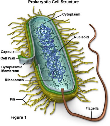
The Cell Envelope comprises the cytoplasmic membrane, cell wall plus an outer membrane, called a capsule (if present):
- Capsule (Only some species of bacteria) - Extra protective - outer capsule composed of polysaccharides (complex carbohydrates). Capsules play a number of roles, but the most important are:
• To keep the bacterium from drying out
• To protect it from phagocytosis (engulfing) by larger microorganisms - a major virulence factor (ability to cause disease) in the major disease-causing bacteria, such as Escherichia coli and Streptococcus pneumoniae. Non-encapsulated mutants of these organisms are avirulent ( i.e. don't cause disease).
- Cell Wall - Each bacterium is enclosed by a rigid cell wall composed of peptidoglycan, a protein-sugar (polysaccharide) molecule.
• Gives the cell its shape and surrounds the cytoplasmic membrane - protecting it from the environment.
• Helps to anchor appendages like the pili (tiny whiskers used to exchange genetic material) and flagella (whip-like flagellae used to propel bacterium in water). These originate in the cytoplasm membrane and protrude through the wall to the outside.
• Wall strength keeps cell from bursting. The primary function of the wall is to protect the cell from internal pressure caused by much higher concentrations of proteins and other molecules inside the cell compared to outside;
• Unique to bacteria is the presence of peptidoglycan. Located immediately outside the cytoplasmic membrane;
• Cell wall composition varies widely amongst bacteria and is one of the most important factors in bacterial species analysis and differentiation. E.g. Cell wall thickness determines whether bacteria is gram-positive or gram-negative (see below)
- Cytoplasmic membrane. Composed of a phospholipid bilayer, similar to a eukaryotic cell membrane, and similarly acts as a permeability barrier for most molecules and serves as a transport zone for molecules into the cell
Gram-positive (Thick-walled)or gram-negative (Thin-walled)? In 1884, Danish physician Hans Christian Gram devised a staining and washing technique to differentiate between bacteria with thick cell walls and those with thin walls. When exposed to a "gram stain", gram-positive bacteria retain the purple color of the stain because the structure of their cell walls traps the dye. In gram-negative bacteria, the cell wall is thin and releases the dye readily when washed with an alcohol or acetone solution.
ACID-FAST bacteria. Won't hold gram stain, so are difficult to define as gram-positive or gram-negative
|
gram-positive |
Gram-negative |
|
Staphylococcus
spp
Clostridium
spp Corynebacterium |
Acinetobacter spp Neisseria Haemophilus spp Bordetella Helicobacter spp (curved rod/helix) Campylobacter Pseudomonas spp Legionella spp Bacteroides
Escheririchia coli Proteus Klebsiella spp Shigella Citrobacter Serratia Morganella Yersinia Enterobacter |
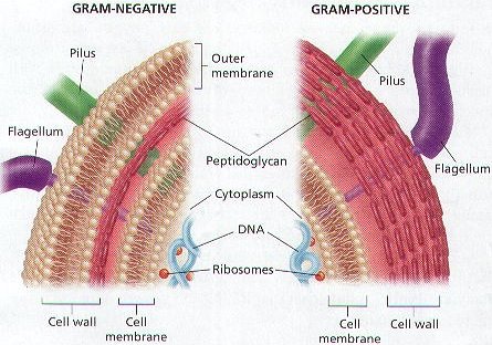
Comparison of BACTERIAL CELL WALLS in Gram-positive and Gram-Negative bacteria
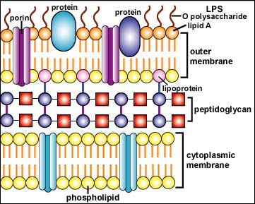
Gram Negative Cell Wall (in detail)
Peptoglycan layer in the cell wall of Gram-negative bacteria is much thinner than the gram-positive bacteria - comprised of only 20% peptidoglycan
Unique to Gram-negative bacteria is an outer membranelayer - high in lipid content, it is formedby lipopolysaccharide (LPS) layer, lipoproteins, proteins, aphospholipid layer and porins:
• Periplasmic space - separates cell plasma membrane from the peptidoglycan layer (contains proteins which destroy potentially dangerous foreign matter present in this space).
• Phospholipid layer - located in inner layer of outer membrane attached by lipoproteins to the exterior peptidoglycan layer;
Many G- bacteria are pathogenic associated with its endotoxin layer
|
The Black Plague wiped out a third of the population of Europe - It was caused by the tiny G- rod, Yersinia pestis |
• PS layer (also known as endotoxin) - this outer layer of the outer membrane is comprised of lipopolysaccharides; their lipid portion is embedded in the outer membrane and is called Lipid A, a toxic substance responsible for most pathogenicity of G- bacteria. Also contributing to toxicity are polysaccharides, called O polysaccharides, extending outward from the bacterial surface.
The highly charged lipopolysaccharides give the G- cell wall an overall negative charge
The center segment, called the core polysaccharide, contains sugars which are highly phosphorylated; it's these phosphate groups which contribute the negative charge.
• Porins -as a phospholipid bilayer, the lipid part of the outer membrane is impermeable to charged molecules. Channels, called porins, span the outer membrane and allow passive transport of solutes, manyions, sugars and amino acids across the outer membrane into and out of the bacterial cytoplasm