Health Happening
The "No-Brainers" for Physical and Mental Health:

 (2) ELIMINATE


Breast Cancer
Research at the American Institute of Cancer Research estimated that about 40% of U.S. breast cancer cases could be prevented by making better lifestyle choices. This figure is likely a low estimate, since the latest paleoanthropological research shows that cancer was virtually nonexistent in humans before poor diet and pollution appeared.
Iron and aluminum toxicity in breast cancer
Genes do not have the last word. Breast cancer risk increases only ~20-30% with a family history of the disease. Mutations of the BRCA1 and BRCA2 genes are said to increase breast cancer risk to 80%. However, it is the expression of your genes that dictates the risk, and not simply their existence. Gene expression can easily be controlled (by whether they are turned on or not) via lifestyle and dietary choices.
| Relative Risk | Factor |
|---|---|
| > 4.0 | Female Age > 65 - although risk increases across all ages until
80; Inherited mutations for breast cancer (BRCA-1 or BRCA-2) 2 or more; 1st-degree relatives diagnosed with BC at an early age; Personal history of BC; High breast tissue density Biopsy confirmed atypical hyperplasia |
| 2.1 - 4.0 | One 1st-degree relative with BC; High dose radiation to chest; High bone density (post-menopausal) |
|
1.1 - 2.0 Factors affecting circulating hormones |
Age >30 years at 1st full-term pregnancy; Menarche before age 12 years; Menopause > 55 years; No full-term pregnancies; Never breast-fed a child; Recent oral contraceptive use; Recent and long-term use of hormone replacement therapy; Postmenopausal obesity |
|
1.1 - 2.0
Other Factors |
Personal history of endometrium, ovary or colon cancer; Alcohol consumption* Tall height; High socio-economic status; Jewish heritage |
* More than 2 alcoholic drinks/day. According to data published in the British Journal of Cancer in 2002, 4% of all breast cancers (~44,000 cases a year) in the United Kingdom are due to alcohol consumption. Could this be due to magnesium depletion in the body consequential to drinking alcohol?
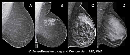
High breast density affects ability to see abnormal tissue on a mammogram. Tumors and calcifications show up white, as does dense breast tissue.
Dense breasts have relatively high amounts of glandular and connective tissue compared to fatty tissue. The diagram below shows increasing levels of glandular / connective tissue density, which can only be seen on mammograms and can not be felt via physical examination. Breasts are classified as dense if they fall into the (C) and (D) classifications:
(A) Almost all fatty breast tissue (10% of women)
(B) Scattered dense glandular / connective tissue (40% of women)
(C) Hetererogeneously dense breast tissue with many areas of dense glandular / connective tissue (40% of women)
(D) Extremely dense tissue (10% of women)
Although often inherited, other factors can influence breast density. Including increasing age, having children and using the estrogen-lowering drug Tamoxifen. Breast tissue density is also associated with having a low body mass and using postmenopausal hormone replacement therapy.
Having dense breast tissue does not increase death rate from breast cancer. Although having high breast tissue increases risk for breast cancer, research shows that a woman with dense breast tissue is no more likely to die from breast cancer than a woman with fatty breast tissue.
Double blind, placebo controlled, randomized study used transdermal Progesterone and ESTRADIOL on 40 premenopausal women undergoing breast surgery for the removal of a lump. The study examined results of two breast biopsies, one at the beginning of the study and another 13 days later.
- Estrogen and the Progesterone did not show up in the serum, but showed up in the breast tissue at over 100% increased levels above placebo. (Chang et al, 1995)
| Method of Measuring Cell Proliferation ▼ | Placebo | Progesterone | ESTRADIOL |
|---|---|---|---|
|
Mitosis per 1000 Cells |
0.51 |
0.17 |
0.83 |
|
PCNA (proliferating cell nuclear antigen) - the most accurate method |
7.8 |
1.9 |
17.4 |
Study concluded that in normal breast tissue:
• Increased ESTRADIOL concentration increased cell proliferation
• Exposure to PROGESTERONE for 10-13 days reduced ESTRADIOL-induced proliferation
Based on PCNA numbers (PCNA presence in actively growing and dividing cells serves as a marker for such cells):
1. Topical Progesterone reduced ▼cell proliferation by 410%
2. Topical ESTRADIOL increased ▲cell proliferation by 223%
Progesterone levels naturally decrease with age. In women this decrease occurs about the age of 35 and men about ten years later. Progesterone balances estrogen, such that an imbalance of estrogen over PROGESTERONE could be responsible for increased cell proliferation in estrogen-sensitive breast cancer.
Chang KJ, Lee TTY , Linares-Cruz G, Fournier S, de Lignieres B (1995) Influences of percutaneous administration of estradiol on human breast epithelial cell cycle in vivo. Fertility and Sterility; 63; 7865-7891. Online link
Ikram Ullah, Govindasamy-Muralidharan Karthik, Amjad Alkodsi, Una Kjällquist, Gustav Stålhammar, John Lövrot, Nelson-Fuentes Martinez, Jens Lagergren, Sampsa Hautaniemi, Johan Hartman, Jonas Bergh (2018) Evolutionary history of metastatic breast cancer reveals minimal seeding from axillary lymph nodes. Journal of Clinical Investigation; DOI: 10.1172/JCI96149